Anterior Shoulder Tendon Anatomy : The Radiology Assistant : Shoulder MR - Anatomy : Majority of anterior shoulder dislocations are due to trauma.
byLawrence Aguirre•
0
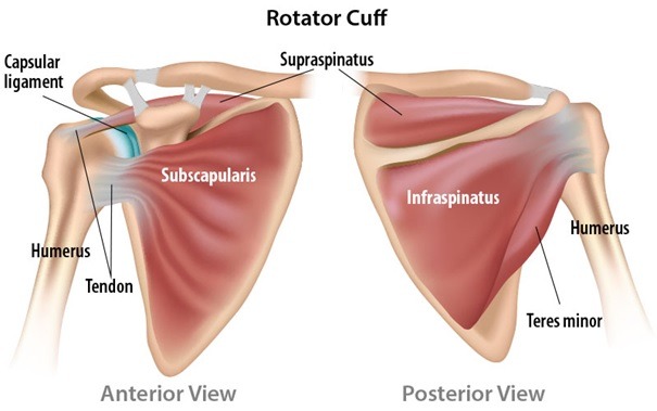
Anterior Shoulder Tendon Anatomy : The Radiology Assistant : Shoulder MR - Anatomy : Majority of anterior shoulder dislocations are due to trauma.. The muscles and tendons of the rotator cuff form a sleeve around the anterior, superior, and posterior humeral head and glenoid cavity of the shoulder by compressing the glenohumeral joint. Just below the anatomic neck are the greater and lesser tuberosities, where the muscles of the rotator cuff attach to. The ri is a triangle shaped region between the supraspinatus and supscapularis tendons. Infraspinatus and teres minor tendon. The pectoralis minor muscle is a small.
Scapula and related structures — the scapula is a relatively large, flat bone located on the posterior thorax (figure 1 and the anterior and posterior portions of the supraspinatus muscle give rise to distinct portions of the supraspinatus tendon. Learn this topic now at kenhub. The tendon crosses anterior to the ankle joint and attaches to the base of the distal phalanx of the great toe. Anterior static shoulder stability is provided by. Biceps tendon is seen anteriosuperiorly & sabscapularis tendon anterior to the scapula at midsuperior head level.
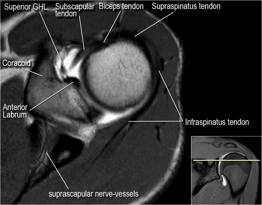
Rotator Cuff Disfunctie - Optimum Zoetermeer from optimumzoetermeer.nl Normal mri anatomy of shoulder joint axial section: The most common shoulder injuries involve the muscles, ligaments, cartilage, and tendons. Mnemonics that can be used to remember the anatomy of the ankle tendons from anterior to posterior as they pass posteriorly to the medial malleolus of the tibia under the flexor retinaculum in the tarsal tunnel include: Irreducible anterior dislocation of the shoulder due to interposition of the long head of bíceps tendón and avulsed part of the labrum, treated arthroscopically; Extends shoulder from flexed position. Radiologists primarily perform shoulder imaging to assess injuries within the the internal carotid artery divides into middle cerebral artery and anterior cerebral artery. Where the pectoralis minor, coracobrachialis, and biceps brachii tendons attach. • pain and/or pop at anterior shoulder but usually not painful after initial event.
And we already have dissected tendons and also dissected nerves and vessels via the deltopectoral approach extending from the coracoid process down to the mid level of the forearm.
Majority of anterior shoulder dislocations are due to trauma. The tendon crosses anterior to the ankle joint and attaches to the base of the distal phalanx of the great toe. Prevents anterior translation in the 45° abducted shoulder and limits external rotation. • review pertinent anatomy and pathology associated with common causes of shoulder pain. Robin smithuis and henk jan van der woude. Shoulder anatomy for ultrasound evaluation. Dynamic anterior shoulder stabilization with the long head of the biceps tendon: The human shoulder is made up of three bones: Shows the cuff muscles as an intermediate signal intensity structures. Ligaments are soft tissue structures that connect bones to bones. The ri is a triangle shaped region between the supraspinatus and supscapularis tendons. Pectoral, anterior shoulder, anterior arm. Infraspinatus and teres minor tendon.
The middle cerebral artery travels to the lateral fissure. Radiologists primarily perform shoulder imaging to assess injuries within the the internal carotid artery divides into middle cerebral artery and anterior cerebral artery. The human shoulder is made up of three bones: Shows the cuff muscles as an intermediate signal intensity structures. Normal mri anatomy of shoulder joint axial section:
The Radiology Assistant : Shoulder MR - Anatomy from radiologyassistant.nl Anterior band of ighl (main restraint). Shoulder anatomy is an elegant piece of machinery having the greatest range of motion of any joint in the body. Bicipital groove is best seen on these. Ligaments are soft tissue structures that connect bones to bones. Normal anatomy, variants and checklist. Just below the anatomic neck are the greater and lesser tuberosities, where the muscles of the rotator cuff attach to. Glenohumeral joint glenohumeral joint the glenohumeral joint is a multiaxial synovial ball and socket joint and involves articulation between the glenoid fossa of the. The middle cerebral artery travels to the lateral fissure.
Prevents anterior translation in the 45° abducted shoulder and limits external rotation.
An image depicting shoulder anatomy can be seen below. Glenohumeral joint glenohumeral joint the glenohumeral joint is a multiaxial synovial ball and socket joint and involves articulation between the glenoid fossa of the. In this episode of eorthopodtv, orthopaedic surgeon randale c. Biceps tendon is seen anteriosuperiorly & sabscapularis tendon anterior to the scapula at midsuperior head level. Specifically, the four rotator cuff muscles include the following Prevents anterior translation in the 45° abducted shoulder and limits external rotation. Important to rule out axillary nerve injury. There are several important ligaments in the shoulder. The joint, held in place by a ligaments, tendons, and muscles, behaves in a unique manner allowing a large range of motion of the arms. The radiocarpal joint is made up of the ___, ___, and. Tendon of the long head of the biceps brachii. Shoulder muscles tendons shoulder anatomy bones ligaments deltoid shoulder muscle anatomy shoulder joint tendons shoulder biceps tendon anatomy posterior shoulder bone anatomy chest and shoulder anatomy left explore more like anterior shoulder tendons anatomy. A cadaveric demonstration video showing anterior shoulder anatomy.
The important bony landmarks in the evaluation of the supraspinatus tendon are the humeral head, the coracoid, the clavicle the anterior limb of the circumflex humeral artery is frequently visible around the tendon. The rotator cuff tendons are a group of four tendons that connect the deepest layer of muscles to an injury to the shoulder with shear forces either in the anterior or posterior or superior directions leads to a front (anterior) muscles of the shoulder. Latarjet procedure performed more commonly than bristow. Specifically, the four rotator cuff muscles include the following Normal mri anatomy of shoulder joint axial section:
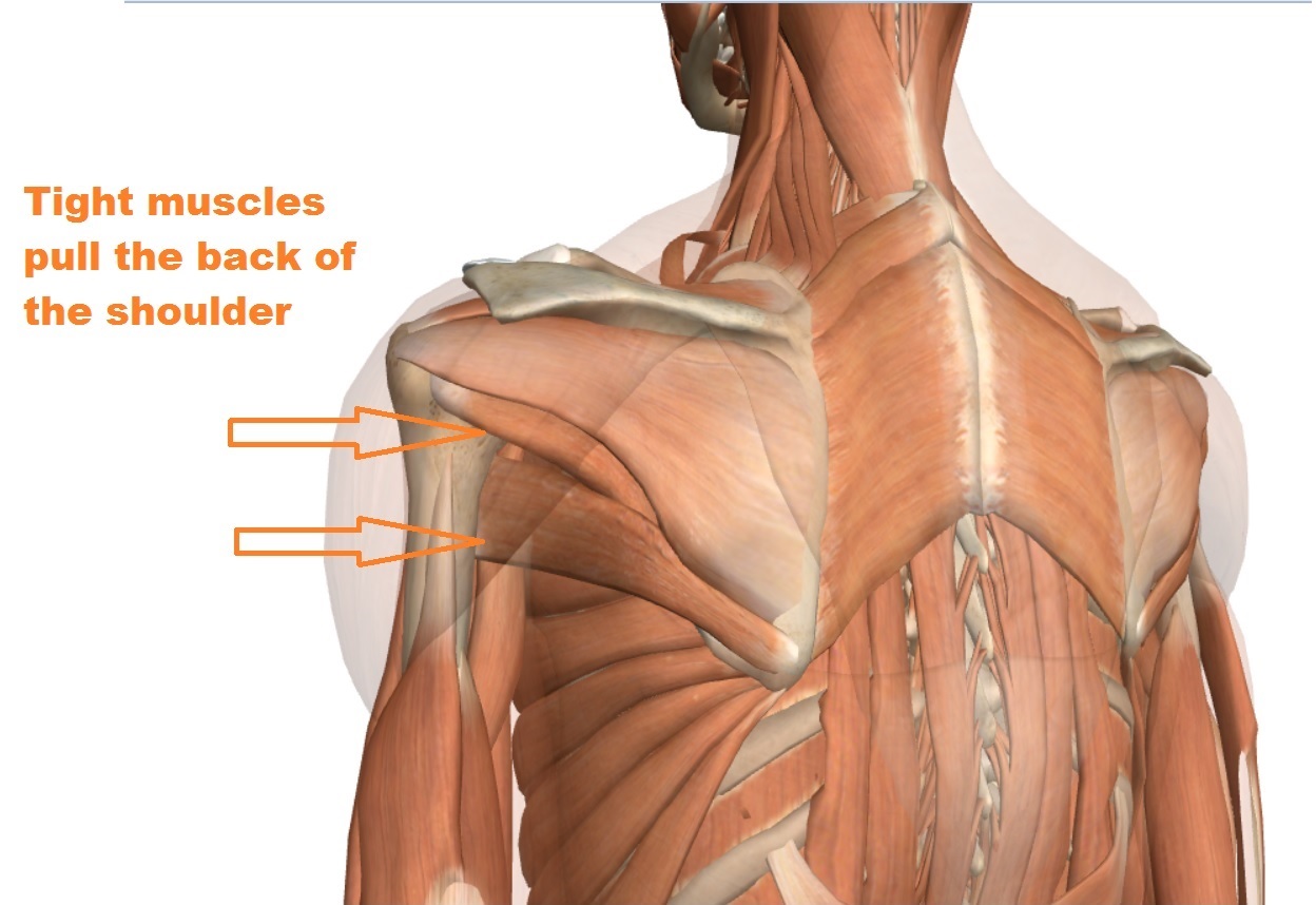
Shoulder Ligament Injuries - Innova Pain ClinicInnova Pain ... from www.innova-pain.com Biceps brachii origin (proximal attachment). Irreducible anterior dislocation of the shoulder due to interposition of the long head of bíceps tendón and avulsed part of the labrum, treated arthroscopically; And we already have dissected tendons and also dissected nerves and vessels via the deltopectoral approach extending from the coracoid process down to the mid level of the forearm. Anterior — the front of the shoulder. Specifically, the four rotator cuff muscles include the following Transfer of coracoid bone with attached conjoined tendon and ca ligament. The shoulder bones in the joint can. Important to rule out axillary nerve injury.
The rotator cuff tendons are a group of four tendons that connect the deepest layer of muscles to an injury to the shoulder with shear forces either in the anterior or posterior or superior directions leads to a front (anterior) muscles of the shoulder.
The pectoralis minor muscle is a small. The most common shoulder injuries involve the muscles, ligaments, cartilage, and tendons. Latarjet procedure performed more commonly than bristow. Tendon of the long head of the biceps brachii. Pectoral, anterior shoulder, anterior arm. Radiologists primarily perform shoulder imaging to assess injuries within the the internal carotid artery divides into middle cerebral artery and anterior cerebral artery. Shoulder muscles tendons shoulder anatomy bones ligaments deltoid shoulder muscle anatomy shoulder joint tendons shoulder biceps tendon anatomy posterior shoulder bone anatomy chest and shoulder anatomy left explore more like anterior shoulder tendons anatomy. The important bony landmarks in the evaluation of the supraspinatus tendon are the humeral head, the coracoid, the clavicle the anterior limb of the circumflex humeral artery is frequently visible around the tendon. Shows the cuff muscles as an intermediate signal intensity structures. Dynamic anterior shoulder stabilization with the long head of the biceps tendon: And we already have dissected tendons and also dissected nerves and vessels via the deltopectoral approach extending from the coracoid process down to the mid level of the forearm. Most common finding is 'military patch' (deltoid) anesthesia. Biceps brachii origin (proximal attachment).
Originates from the medial surface of the fibular shaft shoulder tendon anatomy. This webpage presents the anatomical structures found on shoulder mri.